Ultrasound is a beneficial (and sometimes necessary) procedure for pregnant women. They usually produce two-dimensional black and white scans of the fetus that can reveal important insights such as biological sex, approximate size, abnormalities such as heart problems and clefts. Magnetic resonance imaging (MRI) may be used if your doctor wants to be more viewed. This captures images that can be combined using magnetic fields to create a 3D view of the fetus.
However, MRI is not a catch-all. 3D scans are difficult to interpret in a state sufficient to diagnose the problem, as the visual system is not used to handling 3D volume scans (in other words, the wraparound appearance that also shows the subject’s internal structure). Enter machine learning to help model fetal development more clearly and accurately from the data, but such algorithms were unable to model somewhat random movements and different body types.
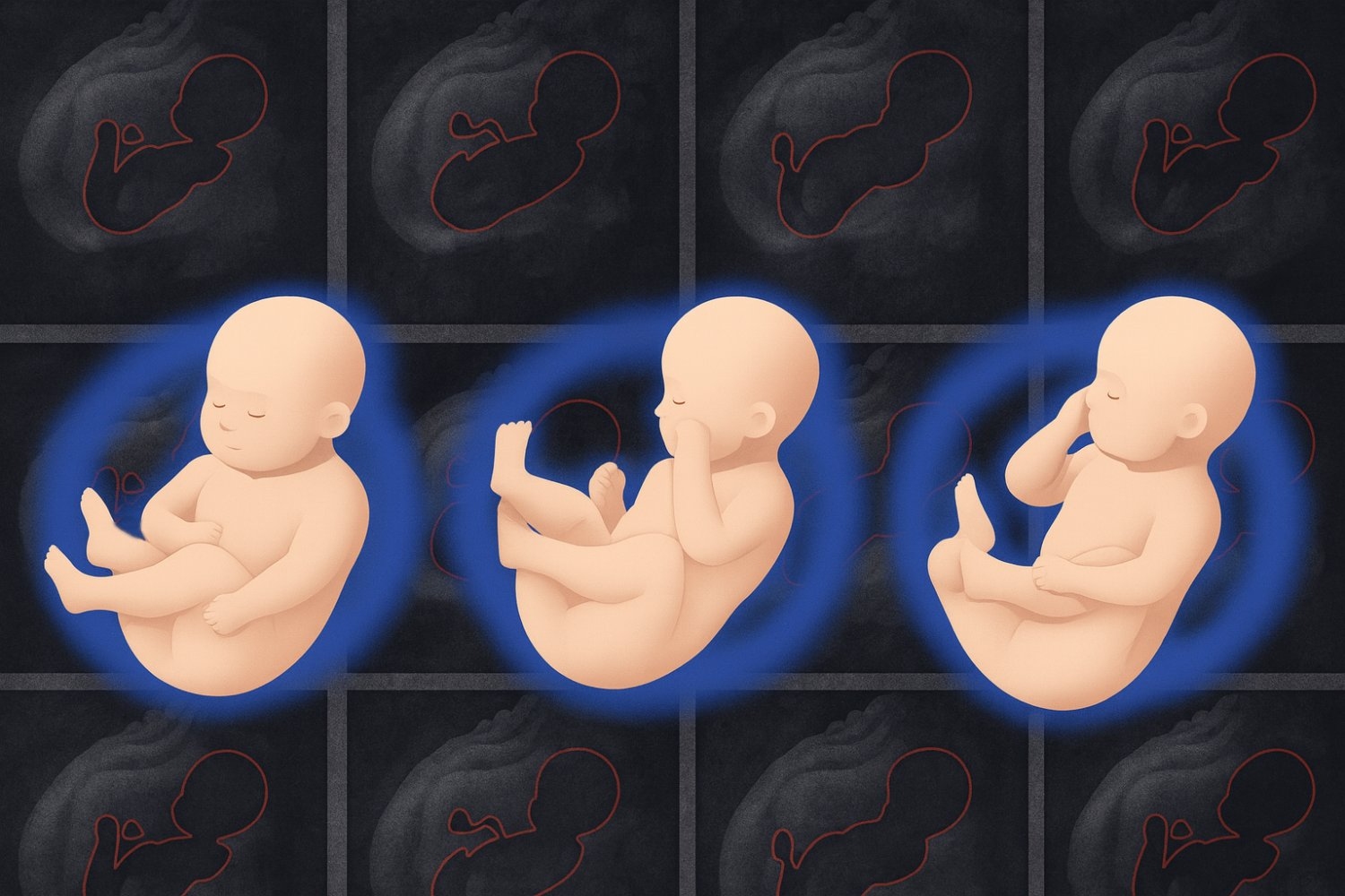
In other words, a new approach called “fetal SMPL” at MIT’s Institute for Computer Science and Artificial Intelligence (CSAIL), Boston Children’s Hospital (BCH), and Harvard Medical School introduced clinicians to detailed photographs of fetal health. As a way to accurately represent the shape and pose of a fetal body, it was adapted from the Skinded Multi-person Linear Model, a 3D model developed in computer graphics. It was developed in computer graphics. Next, the fetal SMPL was trained at 20,000 MRI volumes to predict the fetal position and size and create a sculptural 3D representation. Inside each model there is a skeleton with 23 joints called the “Kinematic Tree,” which the system uses to pose and move like a fetus seen during training.
The extensive, real-world scans learned by the fetal SMPL helped to develop pinpoint accuracy. Imagine stepping into the footsteps of a stranger while blindfolded. Not only does it fit perfectly, but it also correctly guesses which shoes you were wearing. Similarly, the tool closely matched the fetal position and size of the MRI frame, which we had never seen before. Fetal SMPL was only inconsistent by an average of about 3.1 mm, with a gap smaller than a single grain of rice.
This approach allows doctors to accurately measure things like the size of a baby’s head and abdomen and compare these metrics to healthy fetuses of the same age. Fetal SMPL demonstrated clinical potential in early testing, achieving accurate alignment results in small groups of real-world scans.
“It can be difficult to estimate the shape and pose of a fetus, because they are crammed into a solid range of the uterus,” says lead author, student at MIT PhD, and researcher Yingcheng Liu SM ’21, CSAil. “Our approach overcomes this challenge using a system of bones interconnected beneath the surface of a 3D model. This realistically represents the fetal body and its movement. It then relies on a coordinate descent algorithm to make predictions, essentially alternating inferring poses and shapes from tricky data until you find a reliable proponent.”
Inside the uterus
Fetal SMPL was tested with shape and pose accuracy relative to the closest baseline that researchers could find. A system called “Smil” that models infant growth. The baby that emerges from the uterus is larger than the fetus, so the team has reduced these models by 75% to level the arena.
The system exceeded this baseline in a dataset of fetal MRI between 24-37 weeks gestational age taken at Boston Children’s Hospital. The fetal SMPL was able to replicate the actual scan more accurately, as the model lined up closely with the actual MRI.
This method was efficient to arrange models in images, and only required three iterations to arrive at a reasonable alignment. In an experiment that counted the number of false guesses made by fetal SMPL before reaching the final estimate, its accuracy became layered from the fourth step.
Researchers had just begun testing the systems in the real world, and generated similarly accurate models with their first clinical tests. While these results are promising, the team points out that it is necessary to apply the results to larger populations, different ages of gestation and cases of various diseases to better understand the capabilities of the system.
Only deep skin
Liu also points out that only bone-like structures are beneath the skin of the model, so the system can only help analyse the analysis of what the doctor sees on the surface of the fetus. To better monitor the internal health of a baby, such as liver, lungs and muscle development, the team intends to volume the tools and model the internal anatomy of the fetus from scans. Such an upgrade would make the model more human-like, but the current version of fetal SMPL already presents an accurate (and unique) upgrade to 3D fetal health analysis.
“This study introduces methods specifically designed for fetal MRIs that effectively capture fetal movements and enhance the assessment of fetal development and health,” said Kiho IM, Associate Professor of Pediatric Medicine and Staff Scientist in the Department of Neonatal Medicine at BCH’s Center for Fetal Neonatal Science and Developmental Sciences. IM, who was not involved in this paper, adds that the approach “not only improves the diagnostic utility of fetal MRI, but also provides insight into the early functional development of the fetal brain associated with body movement.”
“This study reaches a pioneering milestone by extending the parametric surface human body model of the earliest shape of human life. “This allows for the drought of human shape and movement. This has already proven key to understanding how adult body shapes are related to metabolic conditions and how infant movements are related to neurodevelopmental disorders. Furthermore, fetal models continue the infant model (Smpl) and infant model to continue the continuous shapes of adults (SMPL) and infant models. An unprecedented opportunity to further quantify how human shape growth and movements are affected by various conditions.”
Liu wrote the paper with three CSAIL members. PeiqiWangSM ’22, PhD ’25. MIT PhD student Sebastian Diaz; Senior author Polina Golland, Professor of Electrical Engineering and Computer Science, Chief Researcher at MIT Csail, and Leader of Medical Vision Group. Esra Abaci Turk, an assistant professor of pediatrics in BCH, researcher Benjamin Billot at Inria, and Patricia Ellen Grant, a professor at Harvard Medical School Pediatrics and professor of radiology, are also authors of the paper. This work was supported in part by the National Institutes of Health and the MIT CSAIL-WISTRON program.
The researchers will present their research at the international conference on Medical Imaging Computing and Computer-Assisted Interventions (MICCAI) in September.